
What Happens When the Tissue Biopsy Is Not Enough
In advanced non-small cell lung cancer the starting point for molecular diagnosis is tumour tissue. Once the pathological diagnosis is made, the search for driver changes such as EGFR, ALK, ROS1, BRAF, RET, MET and KRAS determines the choice of treatment directly. The problem is that the sample obtained does not always suffice for those analyses.
A sample can fall short for several reasons: too little tissue from a needle biopsy, a low proportion of tumour cells in the specimen, or DNA or RNA that does not come out at a quality adequate for sequencing. Alongside those, the position of the tumour may make a further procedure risky, or the patient’s general condition may not allow one.
At that point the clinical question is clear: if no further procedure is planned, how is the molecular profile to be obtained? Tumour DNA circulating in the plasma, that is ctDNA, offers a route. The general framework of the method is set out in liquid biopsy for cancer, and exactly what the test measures in ctDNA testing after surgery.
Moving to plasma when the tissue biopsy falls short is common practice. The subject of this article is the evidence that practice rests on and where it stops. The strength of the result obtained varies markedly with the type of change being looked for and with the burden of disease.

Liquid Biopsy or Tissue Biopsy
Whether a test done from blood replaces tissue analysis, and which is chosen in which situation.
What the LC-SCRUM-Liquid Trial Showed
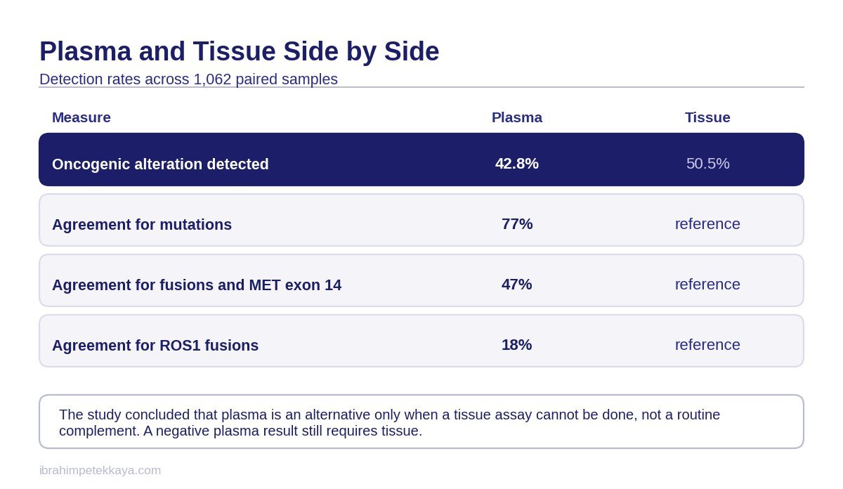
LC-SCRUM-Liquid is a prospective, large-scale concordance study. One thousand one hundred and twelve patients were enrolled; the number with matched blood and tissue samples is 1,062, and the detection rates below belong to that matched group. Stating which set of patients the rates come from is necessary for reading the result correctly.
An oncogenic change was detected in 42.8 per cent of patients with plasma and in 50.5 per cent with tissue. Plasma sequencing therefore missed a proportion of the changes that tissue caught. The gap is about eight percentage points, and it corresponds to whether or not a treatment option can be identified for those patients.
The conclusion the authors themselves drew matters here, and it is often misreported. They did not conclude that plasma complements tissue. What they concluded was that the plasma test can be an option only when tissue analysis cannot be performed because of insufficient DNA and RNA. That is the definition of a fallback route, not of a complementary test.
One favourable finding also came out of the trial. In patients in whom a change was detected in plasma but not in tissue, genotype-matched treatment produced a response rate of 85 per cent and a median progression-free survival of 12.7 months. The changes plasma catches and tissue misses are therefore clinically real; they are not technical noise.
Send Your Reports for Assessment
Share your pathology, imaging and blood results on WhatsApp, or send them by email. Documents are reviewed before the consultation.
Agreement Is Not the Same for Mutations and Fusions
Reporting a single detection rate would be misleading, because agreement with tissue differs markedly with the type of change being sought. That distinction is the piece of information that actually determines how a plasma result is used in the clinic.
For point mutations, agreement with tissue was 77 per cent: 78 per cent for EGFR, 75 per cent for KRAS, 85 per cent for BRAF and 72 per cent for HER2. That level shows plasma to be usable for finding mutations. In a patient in whom a mutation is being sought, a plasma result provides a reasonable basis when tissue is not available.
For fusions and MET exon 14 skipping the same agreement falls to 47 per cent: 46 per cent for ALK, 57 per cent for RET, 66 per cent for MET exon 14 skipping and only 18 per cent for ROS1. An agreement of 18 per cent for ROS1 means that a negative plasma result carries almost no information about that gene.
| Change | Agreement | Note |
|---|---|---|
| Mutations (overall) | 77 per cent | Plasma usable at this level |
| EGFR mutation | 78 per cent | Upper band of the mutation group |
| KRAS mutation | 75 per cent | In line with the mutation group |
| BRAF mutation | 85 per cent | Highest agreement |
| HER2 mutation | 72 per cent | Lower band of the mutation group |
| Fusions and MET exon 14 | 47 per cent | A negative result does not exclude |
| ALK fusion | 46 per cent | Tissue confirmation required |
| RET fusion | 57 per cent | Tissue confirmation required |
| MET exon 14 skipping | 66 per cent | Tissue confirmation advised |
| ROS1 fusion | 18 per cent | A negative plasma result carries no information |
Important: Reporting the detection rate of 42.8 per cent without this distinction makes the result look stronger than it is. In a patient in whom a fusion is being sought, a negative plasma result does not mean there is no fusion.
There is a technical reason for the gap. A point mutation is sought at a single position on the DNA and can be detected even from a small amount of template. A fusion requires the junction of two genes to be captured, and because the DNA fragments in the circulation are short, that junction frequently falls outside the fragment. Analysis performed at the level of RNA in tissue does not carry that limit. The subject is taken up in detail in RNA analysis of tumours.
How Tumour Burden Affects Detectability
One of the things that determines whether a change is detected in plasma is the amount of tumour DNA shed into the circulation. The study by Haseltine and colleagues measured that relationship against tumour volume and showed why a plasma test can come back empty in low-burden disease.
The scope of that study has to be reported accurately. It was not performed during radiotherapy. The patients had stage III and IV disease and had not yet received any treatment at the moment the plasma sample was taken. Because the tumour volumes were contoured in radiation oncology software, the study is from time to time reported as a radiotherapy study; it is not.
The study is single-centre, and of the series of 459 patients enrolled only 110 entered the final analysis. It is therefore a small-scale study rather than a large series. That does not devalue the direction of the finding, but it does prevent a firm threshold being derived from it.
The findings are these: the probability of detecting a mutation in plasma rose, statistically significantly, as total tumour volume increased. The best cut point derived from the data was 64.7 mL, rounded to 65 mL, and above that threshold a multivariable odds ratio of 4.30 was reported.
Bone metastasis was also found to be independently associated with detectability, with a multivariable odds ratio of 2.68. That result is statistically borderline, however, because the lower bound of the confidence interval is 1.06. The direction of the association looks favourable, but in terms of power it is a weak finding.
Important: The 65 mL cut point has not been validated in an external series and appears in no guideline; it is not a clinical decision threshold. The value of the finding is that it explains why a plasma test can come back falsely negative at low tumour burden.
What the Guidelines Say About the Tissue Biopsy
The IASLC consensus document accepts plasma ctDNA as a valid genotyping method in newly diagnosed advanced disease. Starting with plasma at the time of diagnosis is an acceptable approach. The aim there is to shorten the time to starting treatment rather than lose time waiting for the tissue result.
In acquired resistance arising after a targeted drug the position is different: at that stage starting with plasma is the preferred route. If plasma gives no information, the tissue biopsy is repeated. How resistance develops is set out in targeted therapy resistance.
The ESMO guideline on oncogene-addicted metastatic non-small cell lung cancer puts the clearest sentence in this field: cell-free DNA can be used in the search for driver and resistance mutations, but all patients whose blood test comes back negative still require a tissue biopsy. That sentence says directly that plasma does not take the place of tissue.
The ESMO recommendations on ctDNA also state that detection of amplifications and fusions with ctDNA assays is inadequate, and ask for these analyses to be repeated in tissue where possible. That recommendation maps exactly onto the fusion agreement data from LC-SCRUM-Liquid.
Two points need saying openly. There is no ESMO or IASLC liquid biopsy guideline dated 2025 or 2026; the documents that can be cited in this field are the IASLC consensus text and the ESMO guidelines on ctDNA and on oncogene-addicted disease. No specific statement from NCCN documents is reported in this article either, because those documents sit behind a paywall and have not been verified.
When the Pathology Report Alone Is Not Enough
Which further analyses are requested when the pathology report falls short of a treatment decision.
What a Negative Plasma Result Means
No change being detected on a plasma test covers two separate situations: the tumour genuinely not carrying such a change, or the change being present and not caught by the test. The test does not separate the two. A negative result is therefore not an exclusion.
- If the amount of tumour DNA shed into the circulation is low the change can be missed, and that likelihood rises in low-volume disease.
- Fusions and MET exon 14 skipping are structurally harder to capture in the short DNA fragments of the circulation.
- In targets such as ROS1, where agreement is very low, a negative result cannot be used for a decision.
- Amplifications are inadequately detected by plasma assays, and ESMO recommends repeating these analyses in tissue.
- The rule common to every guideline is a single one: on a negative plasma result, go back to tissue.
A plasma test therefore cannot be read as a standalone certificate of result when the tissue biopsy falls short. A positive result directs treatment; a negative result means only an absence of information. That asymmetry is the point that fixes the place of the test in the clinic.
One further detail matters. No guideline says that plasma takes the place of tissue because the tissue was inadequate. What the guidelines say is that plasma can be tried when there is no tissue, and that on a negative result a further procedure is planned in order to go back to tissue. If a further procedure is genuinely not possible, that is discussed openly with the patient.
How the Decision Is Made in Practice
In a patient whose tissue biopsy has fallen short, the order followed is set by the type of target being sought and by whether a further procedure is feasible. The steps below rest on the evidence set out above.
- The pathology report and the sequencing report are read together, and the stage at which the sample fell short is identified.
- The patient’s general condition and the position of the tumour are assessed for the feasibility of a further tissue biopsy.
- If no further procedure can be planned, plasma ctDNA is requested, and it is noted from the outset that the result carries different reliability by type of change.
- If a driver change is detected in plasma, targeted treatment can be started; LC-SCRUM-Liquid reported a response rate of 85 per cent in these patients.
- If plasma is negative the result is not taken as an exclusion; if a fusion or an amplification is being sought, a further procedure for tissue analysis is put on the agenda.
- All the findings are assessed together at the molecular tumour board, and the decision is not tied to any single test.
At which board and on which data the decision is taken is described on the molecular tumour board and treatment pathway pages. Which biomarkers are sought in lung cancer is covered in biomarker testing in lung cancer, and the general treatment framework of the disease on the lung cancer page.
In short, plasma ctDNA is a genuine route to molecular diagnosis when the tissue biopsy falls short, and when it comes back positive it directs treatment. Against that, it does not replace tissue: agreement is low for fusions, a negative result is not an exclusion, and the rule in every guideline is to go back to tissue when plasma is negative.
Send Your Reports for Assessment
Share your pathology, imaging and blood results on WhatsApp, or send them by email. Documents are reviewed before the consultation.
Frequently Asked Questions
What is done when the tissue biopsy comes back inadequate?
First the stage at which the sample fell short is identified. If a further procedure is feasible, the tissue biopsy is repeated. If no procedure can be planned, a plasma ctDNA test is requested, taking into account from the outset that the result carries different reliability by type of change.
Does plasma ctDNA replace the tissue biopsy?
It does not. The conclusion of LC-SCRUM-Liquid itself is that plasma can be an option only when tissue analysis cannot be performed because of insufficient DNA and RNA. The ESMO guideline likewise states that all patients whose blood test comes back negative still require a tissue biopsy.
How good is the agreement between plasma and tissue?
It depends on the type of change. For point mutations agreement is 77 per cent: EGFR 78 per cent, KRAS 75 per cent, BRAF 85 per cent, HER2 72 per cent. For fusions and MET exon 14 skipping it falls to 47 per cent, and for ROS1 it is only 18 per cent.
Does a negative plasma result mean there is no target?
No. A negative result means either that the change is absent or that it is present and was not caught; the test does not separate the two. In fusion targets such as ROS1 and ALK in particular, a negative plasma result is not an exclusion and tissue has to be revisited.
Does tumour burden affect the plasma result?
It does. In a single-centre study of 110 patients the probability of detecting a mutation in plasma rose significantly with tumour volume, and above the 65 mL threshold derived from the data the odds ratio was 4.30. That threshold has not been validated in an external series and is not a decision threshold.
Is a change seen in plasma but not in tissue real?
In LC-SCRUM-Liquid, genotype-matched treatment in these patients produced a response rate of 85 per cent and a median progression-free survival of 12.7 months. These changes are therefore clinically real and do respond to treatment.
Which test is used first in resistance?
In acquired resistance arising after a targeted drug, the IASLC consensus document prefers starting with plasma. If plasma gives no information, the tissue biopsy is repeated. At the time of a new diagnosis, starting with plasma is an acceptable approach.
Related Articles
Related Pages
Lung Cancer
Open the Page
Comprehensive Genomic Profiling
Open the Page
Molecular Tumour Board
Open the Page
Targeted Therapy
Open the PageSources
- Sugimoto A, Matsumoto S, Udagawa H et al. A Large-Scale Prospective Concordance Study of Plasma- and Tissue-Based Next-Generation Targeted Sequencing for Advanced Non-Small Cell Lung Cancer (LC-SCRUM-Liquid). Clin Cancer Res 2023;29(8):1506-1514. Link
- Haseltine JM, Offin M, Flynn JR et al. Tumor volume as a predictor of cell free DNA mutation detection in advanced non-small cell lung cancer. Transl Lung Cancer Res 2022;11(8):1578-1590. Link
- Rolfo C, Mack P, Scagliotti GV et al. Liquid Biopsy for Advanced NSCLC: A Consensus Statement From the IASLC. J Thorac Oncol 2021;16(10):1647-1662. Link
- Pascual J, Attard G, Bidard FC et al. ESMO recommendations on the use of circulating tumour DNA assays for patients with cancer. Ann Oncol 2022;33(8):750-768. Link
- Hendriks LE, Kerr KM, Menis J et al. Oncogene-addicted metastatic non-small-cell lung cancer: ESMO Clinical Practice Guideline. Ann Oncol 2023;34(4):339-357. Link