
Gastrointestinal Cancers Covered
Gastrointestinal cancers arise in the digestive tract from mouth to anus and in the liver, bile ducts and pancreas; according to IARC, colorectal, stomach, liver and oesophageal cancers are all among the 10 most common cancers worldwide. Colorectal, pancreatic and stomach cancer have their own pages; the other organs are covered briefly here.
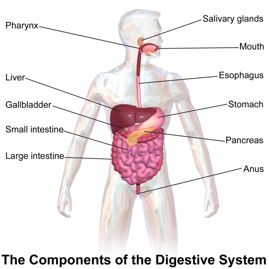
| Organ | Common symptom | Page |
|---|---|---|
| Colon and rectum | Blood in the stool, change in bowel habit, anaemia | Colorectal cancer |
| Stomach | Early fullness, upper abdominal pain, weight loss, anaemia | Stomach cancer |
| Pancreas | Abdominal pain radiating to the back, jaundice, new-onset diabetes | Pancreatic cancer |
| Oesophagus | Difficulty swallowing, pain behind the breastbone, weight loss | This page |
| Liver (hepatocellular carcinoma) | Right upper abdominal pain, fluid in the abdomen, jaundice; usually on a background of cirrhosis | This page |
| Bile ducts and gallbladder | Painless jaundice, itching, dark urine | This page |
| Neuroendocrine tumours and GIST | Often found by chance; flushing, diarrhoea or bleeding | This page and sarcomas |
In locally advanced oesophageal cancer, chemoradiotherapy or chemotherapy before surgery is followed by surgery and immunotherapy. In hepatocellular carcinoma drug treatment rests on immunotherapy-based combinations such as atezolizumab with bevacizumab or durvalumab with tremelimumab; in bile duct cancers immunotherapy is added to chemotherapy and targets such as FGFR2 and IDH1 are sought. GIST is treated with targeted tablets such as imatinib.
Shared Risk Factors
The shared risk factors for gastrointestinal cancers are smoking, alcohol, excess weight, inactivity, processed meat and a diet low in fibre; these act together in colorectal, stomach, oesophageal and pancreatic cancer. Organ-specific factors are Helicobacter pylori in stomach cancer, hepatitis B and C with cirrhosis in liver cancer, and long-standing reflux in oesophageal cancer.
Hereditary syndromes account for about 5 per cent of cases. Lynch syndrome raises the risk of colorectal, stomach and womb cancer, and a BRCA2 mutation raises the risk of pancreatic cancer. Colorectal cancer under 50 in a relative, several affected relatives or an MSI-high tumour are reasons for hereditary assessment.
Anyone with blood in the stool, a change in bowel habit lasting more than six weeks, difficulty swallowing, jaundice, unexplained weight loss or iron-deficiency anaemia should see a physician. The Turkish Ministry of Health screening programme offers a stool test for hidden blood every two years and a colonoscopy every 10 years between the ages of 50 and 70.
Diagnosis and Staging
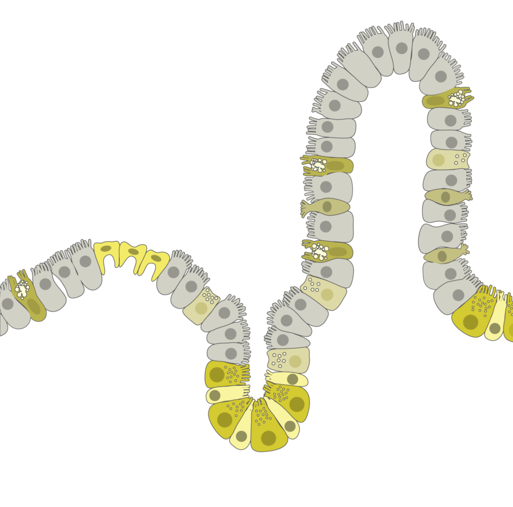
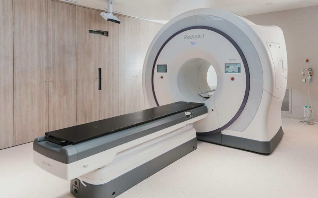
Gastrointestinal cancers are diagnosed by pathology examination of tissue taken at endoscopy (colonoscopy, gastroscopy) or by image-guided biopsy. Staging uses CT of the chest and abdomen, with PET-CT and liver MRI where needed; in rectal cancer a pelvic MRI completes staging. For a remote review, the endoscopy report, the pathology report and the staging CT in DICOM format are the documents that matter most.
Staging follows the TNM system: depth of the tumour in the organ wall (T), lymph node involvement (N) and distant metastasis (M). In stage 1 and 2 surgery is the main treatment; in stage 3 systemic therapy is added to surgery; in stage 4 systemic therapy leads. In colorectal cancer with a few metastases confined to the liver, surgery and systemic therapy are planned together with the surgical team, in Izmir or at the home centre.
Tumour markers (CEA, CA 19-9, AFP) do not make a diagnosis; they follow response to treatment and detect recurrence. CEA is used in colorectal cancer, CA 19-9 in pancreatic and bile duct cancer and AFP in liver cancer; because they also rise in benign conditions, the result is always read alongside imaging. Treatment choice is determined by the physician according to stage, pathology and molecular findings.
Systemic Treatment Options
Chemotherapy is the backbone of systemic therapy in gastrointestinal cancers; fluoropyrimidines (5-fluorouracil or capecitabine), oxaliplatin, irinotecan, taxanes and gemcitabine are used in different combinations. Administration and side effects are described on the chemotherapy page.
- Chemotherapy
- FOLFOX or CAPOX in colorectal cancer, FOLFIRINOX or gemcitabine with nab-paclitaxel in pancreatic cancer and the FLOT regimen in stomach and oesophageal cancer are the standard regimens. Cycles are given every two or three weeks, mostly into a vein; a regimen started in Izmir can be continued at the home centre with a written protocol if the home team agrees.
- Targeted therapy
- EGFR antibodies (cetuximab, panitumumab) in RAS wild-type colorectal cancer, bevacizumab, trastuzumab in HER2-positive stomach cancer, encorafenib in BRAF V600E-mutated colorectal cancer and zolbetuximab in claudin 18.2-positive stomach cancer. Details: targeted therapy.
- Immunotherapy
- In MSI-high tumours pembrolizumab or nivolumab is effective alone in the first line; in stomach, oesophageal and liver cancer it is added to chemotherapy according to PD-L1. Details: immunotherapy.
Nutritional support is a separate heading in these cancers; weight loss is common because of difficulty swallowing, early fullness or pancreatic enzyme deficiency. Dietitian assessment, enzyme replacement and, where needed, tube (enteral) or intravenous (parenteral) feeding are part of the plan; reported dietary requirements are taken into account.
Please note: the information on this page describes general principles; it does not replace a medical consultation or an individual treatment plan. As of 21 September 2026, licensing and reimbursement status in Türkiye differ by drug and are discussed at the consultation.
Molecular Tests
Four biomarkers directly change treatment in gastrointestinal cancers: MSI/MMR, HER2, RAS with BRAF, and NTRK; the ESMO and NCCN guidelines define which is tested in which cancer. MSI/MMR (microsatellite instability or DNA mismatch repair deficiency) is tested at diagnosis in every colorectal and stomach cancer; if high, immunotherapy becomes an option and Lynch syndrome is investigated. RAS and BRAF determine whether an EGFR antibody will work in metastatic colorectal cancer.
HER2 is tested in metastatic stomach and oesophageal cancer for the trastuzumab decision, and in RAS wild-type colorectal cancer for HER2-targeted therapy. NTRK fusions are rare (under 1 per cent); when present, larotrectinib or entrectinib is used.
In metastatic disease these tests are usually run together as comprehensive genomic profiling, which can be ordered on an existing biopsy block sent from another country. A test result does not always change treatment; but in metastatic colorectal cancer targeted therapy is not chosen without the RAS, BRAF and MSI results.
Frequently Asked Questions
What is gastrointestinal cancer?
Gastrointestinal cancer is the collective name for cancers arising in the digestive tract (oesophagus, stomach, small bowel, colon, rectum, anus) and in the organs attached to it, the liver, bile ducts and pancreas. Together these cancers account for about a quarter of all cancers. Their treatment differs by organ, but the biomarker logic and the chemotherapy drugs are largely shared, which is why a medical oncologist assesses them as one group.
When should I see an oncologist rather than a gastroenterologist?
For first symptoms such as blood in the stool, difficulty swallowing, abdominal pain or weight loss, the right first contact is a gastroenterologist, because the diagnosis is made by endoscopy and biopsy. A medical oncologist is consulted once the pathology report confirms cancer; the oncologist completes staging, agrees the treatment sequence with the surgical team and plans drug treatment. Where surgery is planned, an oncology opinion beforehand can change the order of treatment in some cancers, and this review can be done remotely from the existing reports.
What does MSI-high mean?
MSI-high (microsatellite instability-high) means that the tumour's system for repairing DNA errors is defective, so the tumour has accumulated a large number of mutations; the pathology report may also record it as mismatch repair deficiency (dMMR). About 15 per cent of colorectal cancers and 8 to 10 per cent of stomach cancers are in this group. MSI-high tumours respond well to immunotherapy; in metastatic disease pembrolizumab proved superior to chemotherapy in the first line (KEYNOTE-177). The result is also a reason for hereditary testing for Lynch syndrome.
Are oesophageal and liver cancers assessed?
Yes. Oesophageal cancer, liver cancer (hepatocellular carcinoma), bile duct, gallbladder, small bowel and anal cancers, neuroendocrine tumours and GIST are all assessed within medical oncology. These cancers do not have separate pages, but their treatment principles are summarised on this page. In liver cancer the degree of cirrhosis, and in oesophageal cancer the nutritional state, shape the plan; both need joint decisions with surgery and radiation oncology, and the existing reports can be reviewed before any travel.
What do CEA and CA 19-9 show?
CEA and CA 19-9 are proteins that some tumours release into the blood; CEA is measured in colorectal cancer and CA 19-9 in pancreatic and bile duct cancer to follow response to treatment and to detect recurrence. They are not used to make a diagnosis, and they can also rise in benign conditions such as smoking, pancreatitis, cirrhosis or bile duct obstruction. A falling value during treatment suggests response and a rising value during follow-up raises suspicion of recurrence; the decision is always made together with imaging.
Related Pages
Colorectal Cancer
Open the Page
Pancreatic Cancer
Open the Page
Stomach Cancer
Open the Page
Chemotherapy
Open the Page
Immunotherapy
Open the Page
Comprehensive Genomic Profiling
Open the Page
International Patients
Open the Page
Cancer Types
Open the PageSources and Guidelines
- European Society for Medical Oncology. ESMO Clinical Practice Guidelines: Gastrointestinal Cancers. Link
- Cervantes A et al. Metastatic colorectal cancer: ESMO Clinical Practice Guideline. Ann Oncol 2023;34(1):10-32. Link
- Lordick F et al. Gastric cancer: ESMO Clinical Practice Guideline. Ann Oncol 2022;33(10):1005-1020. Link
- Conroy T et al. Pancreatic cancer: ESMO Clinical Practice Guideline. Ann Oncol 2023;34(11):987-1002. Link
- National Comprehensive Cancer Network. NCCN Clinical Practice Guidelines in Oncology: Colon Cancer. Link
- National Comprehensive Cancer Network. NCCN Clinical Practice Guidelines in Oncology: Gastric Cancer. Link
- André T et al. Pembrolizumab in microsatellite-instability-high advanced colorectal cancer (KEYNOTE-177). N Engl J Med 2020;383:2207-2218. Link
- International Agency for Research on Cancer (WHO). GLOBOCAN 2022, Global Cancer Observatory. Link
- Republic of Türkiye Ministry of Health, General Directorate of Public Health. Cancer statistics and national screening standards. Link
- Turkish Society of Medical Oncology. Guidelines and patient information resources. Link