
What DNA Shows and What RNA Shows
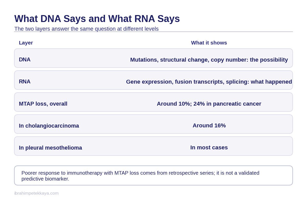
DNA sequencing shows possibility. Mutations, structural changes and copy number changes are read at the level of DNA, and they say what has gone wrong in the genetic code of the cell. But not everything that has gone wrong in the code has to have a counterpart in the cell.
RNA shows what is actually happening. Gene expression, the splicing pattern, fusion transcripts and pathway activity are read at the level of RNA. This is where it becomes clear which genes the cell is running at that moment and which protein blueprint it is producing.
The practical meaning of that distinction is this: cancer is not only a genomic error but a dynamic process. Two tumours carrying the same mutation can behave differently, because which pathways are switched on depends on the level of expression. DNA cannot measure that dynamic.
What a report says at each layer is covered in how to read an NGS report, and the testing process itself on the comprehensive genomic profiling page. For the concept of a driver change, see driver mutation or passenger.

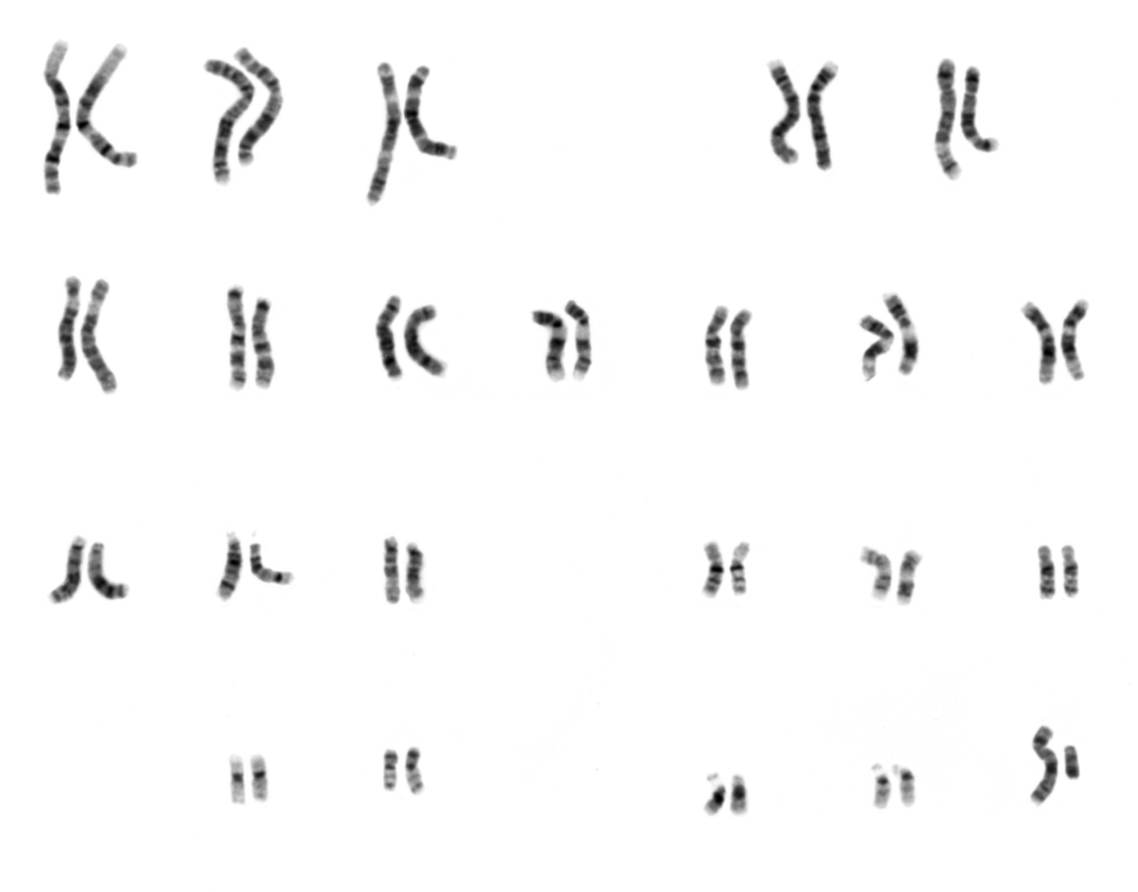
How an NGS Report Is Produced Step by Step
The stages a next-generation sequencing report goes through before it is issued.
What RNA Analysis Adds in the Clinic
The clinical contribution of RNA takes concrete form under three headings, and each of them arises from a structural limit of DNA.
The first is fusions. Because RNA reads the expressed and spliced transcript, it can capture fusions whose breakpoint falls in a large or repetitive intronic region. A DNA capture method can miss the breakpoint in those regions. NTRK and ROS1 fusions are the best-known examples of this.
The second is confirmation. Detecting a fusion at the level of DNA does not show that the fusion is genuinely expressed or that it is in frame. RNA answers both questions directly: is the transcript there, and is the reading frame preserved. That is what determines whether a targeted drug is genuinely meaningful.
The third is exon skipping. Skipping of exon 14 of the MET gene is read directly at the level of RNA. In DNA it has to be inferred indirectly from a change in the splice region. A direct reading carries fewer assumptions than an inference.
What these headings mean clinically in lung cancer is taken up in biomarker testing in lung cancer.
RNA Analysis Is Not More Accurate, It Is Complementary
There is a frequent over-generalisation at this point that needs correcting. RNA is not a wholly more accurate method than DNA. The accurate word is complementary: each method reads a layer the other cannot see.
RNA analysis has its own mode of failure. It depends on the quality of the RNA obtained from formalin-fixed, paraffin-embedded tissue. RNA is more prone to degradation in paraffin blocks, and when the quality is insufficient the test either gives no result or gives a false negative. That is a real limit met in practice, not a theoretical one.
The decision is therefore built like this: DNA and RNA are placed beside one another rather than in place of one another. DNA carries the mutation and copy number side, RNA the expression and fusion side. Reading the two together produces a more reliable picture than the result of either method alone.
Doing that reading is the work of a board rather than of one doctor. How much weight each layer is given is set out on the molecular tumour board page.
Send Your Reports for Assessment
Share your pathology, imaging and blood results on WhatsApp, or send them by email. Documents are reviewed before the consultation.
What MTAP Loss Is and How Often It Occurs
The MTAP gene sits in the 9p21 region, and deletions there remove it together with CDKN2A. MTAP loss therefore rarely comes alone: it is part of a region that is deleted along with neighbouring tumour suppressor genes.
Frequency cannot be given as a single number. Across cancers in general it is seen at a level of roughly 10 per cent; in pancreatic cancer that figure rises to about 24 per cent, and in cholangiocarcinoma to about 16 per cent. In pleural mesothelioma, MTAP loss is present in the large majority of cases.
This distribution explains why MTAP loss is discussed most in pancreatic and biliary tract tumours. In diseases where the frequency is high, any treatment target arising from the loss would affect a large number of patients. The wider framework of the disease is set out on the pancreatic cancer page.
| Disease | Frequency |
|---|---|
| Cancers in general | Roughly 10 per cent |
| Pancreatic cancer | About 24 per cent |
| Cholangiocarcinoma | About 16 per cent |
| Pleural mesothelioma | The large majority of cases |
Does Seeing the Deletion Prove the Loss
Detecting a deletion at the level of DNA does not automatically mean the protein is genuinely not being produced at all. That is what creates the problem of disagreement between methods of measurement.
MTAP loss can be assessed by three separate methods: immunohistochemistry at the level of protein, FISH at the level of the chromosome, and next-generation sequencing at the level of DNA. These three do not always give the same answer. The literature openly calls for the methods to be harmonised.
No percentage agreement is given here, because there is no reliable measurement that reduces agreement between the methods to a single number. In the same way, no prospective study comparing DNA and RNA methods for MTAP loss exists.
What that means in the clinic is this: if a report states MTAP loss, the method by which it was detected should be asked for. If a drug choice is to rest on that information, the result of a single method may not be accepted as sufficient. Questions of VUS and classification are taken up in what does VUS mean on an NGS report.
Not Every Mutation Carries the Same Clinical Weight
How the changes seen on a genetic report are classified by clinical importance.
Three Claims, Three Different Strengths of Evidence
Three separate claims about MTAP loss are in circulation. They are not of equal strength and have to be read one at a time.
The first is poor prognosis, and it is the best supported. It has been found consistently across independent patient groups. In cholangiocarcinoma an adjusted hazard ratio of 3.95 has been reported for overall survival. There is one caveat, however: in the large majority of cases MTAP is deleted together with CDKN2A and CDKN2B, so the effect cannot be attributed to MTAP alone.
The second is a reduced response to checkpoint inhibitors, and it is the weakest of the three. MTAP loss has been associated with worse outcomes in retrospective series; it is not a validated predictive biomarker. A study in the same field also argues that the immunologically quiet picture in the 9p21 region arises not from MTAP but from loss of interferon-epsilon.
The third is that the tumour microenvironment is immunologically quiet. This claim is supported, but the effect should be attributed to co-deletion of the 9p21 region rather than to MTAP. For pancreatic cancer the description immune-excluded and fibrotic is more accurate than cold, because the problem is not that immune cells are absent but that they cannot get into the tumour.
| Claim | Status |
|---|---|
| Poor prognosis | Supported in independent groups, with the CDKN2A co-deletion caveat |
| Reduced checkpoint response | Retrospective series only; not a predictive biomarker |
| Immunologically quiet microenvironment | Supported; attributed to 9p21 co-deletion |
Important: MTAP loss is not today a validated predictive biomarker that can be used for an immunotherapy decision. Of these three claims, only the prognosis heading has been found consistent across independent groups.
How Synthetic Lethality Is Built
The MTAP gene works in the methionine salvage pathway. When the gene is deleted that pathway breaks, and an intermediate called MTA accumulates inside the cell. The crucial point of the mechanism is what that accumulation does.
The accumulated MTA competes with SAM at the substrate site of the PRMT5 enzyme and partly inhibits it. Care is needed here: MTA activates nothing. What it sets up is a partial inhibition. The enzyme does not stop altogether but is left running more slowly than normal.
The consequence is that the cell becomes hypersensitive to the same pathway being inhibited once more. Inhibiting an already partly inhibited enzyme a little further kills a cell that has lost MTAP while leaving a cell with intact MTAP unharmed. That is what is meant by synthetic lethality.
It would be wrong here to describe PRMT5 simply as an essential gene. What the drugs do is not to switch off an essential gene but to add pressure to an enzyme that is already partly inhibited. The therapeutic window arises from exactly that difference.
The link to the RNA side is this: whether a deletion has genuinely turned into a loss at protein level is judged by looking at the expression layer. MTAP is therefore the concrete example of why DNA and RNA need to be read together.
Where the Drugs Stand
Two classes of drug exploit this mechanism. The first are MTA-cooperative PRMT5 inhibitors, designed to bind more strongly in a cell in which MTA has accumulated. The second are MAT2A inhibitors, which press on the same pathway by reducing the production of SAM.
To see why that design was necessary, look at what happened to the first generation. Non-selective PRMT5 inhibitors failed because of dose-limiting haematological toxicity. The MTA-cooperative design was developed precisely to solve that problem.
Today’s position has to be stated plainly: no PRMT5 or MAT2A inhibitor is approved anywhere in the world, in any indication. A number of programmes have been stopped: TNG908, GSK3326595, PF-06939999 and AG-270 were discontinued, and S095033 was withdrawn.
On the continuing side, randomised phase 2 and phase 3 trials of an MTA-cooperative PRMT5 inhibitor are running in pancreatic and lung cancer. Early-phase activity has been described as promising, but because there is no peer-reviewed publication of clinical efficacy for these agents, no response rate is given here.
What there is to do today in a patient found to have MTAP loss is therefore not to look for an approved drug but to assess whether a suitable clinical trial exists. The results of biomarker-driven prospective trials are still awaited. Routes of access are described in early access programme and clinical trials.
The general framework for reading reports and reaching decisions is covered on the precision oncology page, and how targeted drugs work on the targeted therapy page.
Finding a Mutation Does Not Mean There Is a Treatment
Why seeing a change on a genetic report does not translate directly into a treatment.
Send Your Reports for Assessment
Share your pathology, imaging and blood results on WhatsApp, or send them by email. Documents are reviewed before the consultation.
Frequently Asked Questions
Is RNA analysis more accurate than DNA sequencing?
No. The two methods are complementary; neither takes the place of the other. DNA reads the mutation, structural change and copy number side; RNA reads gene expression, the splicing pattern and fusion transcripts. The limit of RNA analysis is that RNA of sufficient quality cannot always be obtained from a paraffin block.
In which situations does RNA analysis make a difference?
In fusions whose breakpoint falls in a large or repetitive intronic region, of which NTRK and ROS1 are the known examples; in confirming that a fusion is genuinely expressed and in frame; and in reading MET exon 14 skipping directly rather than by inference.
How often does MTAP loss occur?
Roughly 10 per cent across cancers in general. About 24 per cent in pancreatic cancer and about 16 per cent in cholangiocarcinoma; in pleural mesothelioma it is present in the large majority of cases. MTAP sits in the 9p21 region and is deleted together with CDKN2A.
Does MTAP loss mean a poor prognosis?
Its association with poor prognosis is supported across independent patient groups, and an adjusted hazard ratio of 3.95 has been reported for overall survival in cholangiocarcinoma. But because MTAP is deleted together with CDKN2A and CDKN2B in the large majority of cases, the effect cannot be attributed to MTAP alone.
Does MTAP loss change an immunotherapy decision?
It does not. MTAP loss has been associated with worse outcomes in retrospective series; it is not a validated predictive biomarker. One study also argues that the immunologically quiet picture in the 9p21 region arises from loss of interferon-epsilon.
How does synthetic lethality work here?
When MTAP is deleted the methionine salvage pathway breaks and MTA accumulates. The accumulated MTA competes with SAM at the substrate site of the PRMT5 enzyme and partly inhibits it. The cell thereby becomes hypersensitive to the same pathway being inhibited once more.
Can a PRMT5 or MAT2A inhibitor be used?
No. No drug of this class is approved anywhere in the world, in any indication. TNG908, GSK3326595, PF-06939999 and AG-270 were discontinued and S095033 was withdrawn. Randomised trials of an MTA-cooperative PRMT5 inhibitor are running in pancreatic and lung cancer.
Related Articles

Related Pages
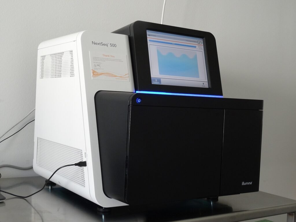
Comprehensive Genomic Profiling
Open the Page
Precision Oncology
Open the Page
Molecular Tumour Board
Open the Page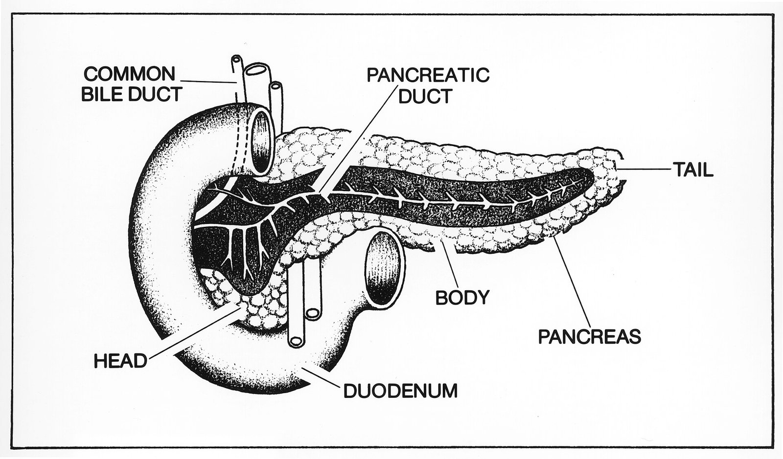
Pancreatic Cancer
Open the PageSources
- Rodon J, Johnson ML, George B et al. MTAP Deletion in Oncogenesis: A Synthetic Lethality Scenario. Cancer Res 2026;86(7):1558-1569. Link
- Arter ZL, Shim JSG, Park C et al. MTAP loss in non-small-cell lung cancer and beyond: Therapeutic targeting of the MAT2A-PRMT5 axis. Cancer Treat Rev 2026;150:103204. Link
- Bragança Xavier C, Shah PA, Yakimova D et al. Convergence of KRAS Mutations and MTAP Loss in Pancreatic Cancer. Clin Cancer Res 2026;32(17):3921-3933. Link
- Lyon J, Osataphan S, Birda V et al. Prognostic Role of MTAP Loss in Cholangiocarcinoma. JCO Precis Oncol 2025;9:e2400842. Link
- Brune MM, Savic Prince S, Vlajnic T et al. MTAP as an emerging biomarker in thoracic malignancies. Lung Cancer 2024;197:107963. Link
- Marjon K, Cameron MJ, Quang P et al. MTAP Deletions in Cancer Create Vulnerability to Targeting of the MAT2A/PRMT5/RIOK1 Axis. Cell Rep 2016;15(3):574-587. Link
- Ju S, Cui Z, Hong Y et al. Detection of multiple types of cancer driver mutations using targeted RNA sequencing in non-small cell lung cancer. Cancer 2023;129(15):2422-2430. Link