
What Biomarker Testing in Lung Cancer Looks For
Guidelines require certain genetic alterations to be looked for in advanced non-small-cell lung cancer. Each one matches a drug developed against it.
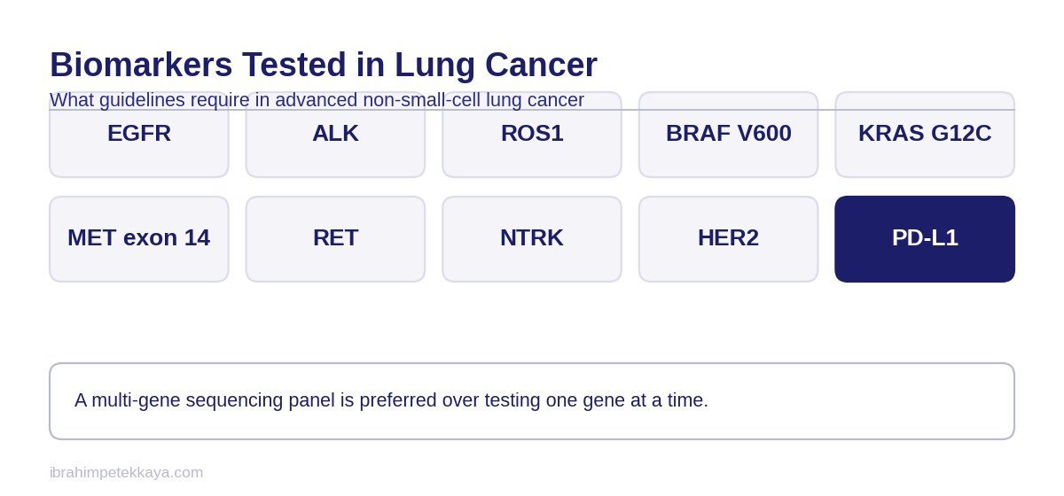
The European guideline recommends testing for EGFR, ALK, ROS1, BRAF V600, NTRK, MET exon 14 skipping, MET amplification, RET, KRAS G12C and HER2 alterations. It also states that PD-L1 should be determined systematically in all patients with advanced disease; that measurement informs the immunotherapy decision.
What these genes share is that each is tied to a drug decision: if an alteration is found, treatment may change. Genes left off the panel do not change a decision today, and the list widens with new approvals.
In the joint guideline from the American pathology organisations, EGFR, ALK and ROS1 testing is mandatory in all lung adenocarcinomas irrespective of clinical characteristics. Other genes are recommended as part of broader panels rather than as stand-alone tests. That document dates from 2018 and predates the approvals for RET, MET exon 14, NTRK and KRAS G12C, so the current gene list should be read from the European guideline.
Why a Panel Rather Than Single Genes
Testing genes one at a time exhausts tissue and time together. Each test takes a section, and after several in sequence the block may hold too few cells. Turnaround adds up as well, pushing treatment back by weeks. A panel reads the same sample once and avoids both losses.
The second reason is biological. The drivers sought on a panel rarely occur together in one tumour, so finding one lowers the likelihood of the others. Since which will be found cannot be known in advance, any sequence usually starts with the least likely gene.
Coverage holds a further detail. ALK, ROS1, RET and NTRK alterations usually appear as fusions, and where the breakpoint falls in a non-coding part of the gene they cannot be detected reliably on a DNA panel. Fusions are therefore also pursued at RNA level, as described under RNA analysis of tumours.
When It Is Requested
The test is requested before treatment starts. That simple rule is often missed in practice, and the consequences are serious.
In the Aggarwal real-world cohort of patients with advanced non-squamous lung cancer, 20 per cent did not have molecular results available when first-line treatment began. Patients whose results were available in time had longer overall survival. This is real-world data and the gap may largely reflect a difference in the patients’ general condition: those started urgently are by definition the ones deteriorating fastest. In the same study, testing tissue and plasma concurrently roughly doubled the odds of the result arriving in time.
Important: Treatment started before results are back can expose the patient to a less effective regimen and disturb the sequence in which targeted drugs are used. Unless the situation is urgent, waiting for the result is the rule and starting is the exception.

Genetic Analysis Before Treatment in Lung Cancer
Identifying the alteration the tumour carries directly changes which treatment is chosen at the first step.
Send Your Reports for Assessment
Share your pathology, imaging and blood results on WhatsApp, or send them by email. Documents are reviewed before the consultation.
Tissue or Blood?
The two methods complement rather than replace each other. Tissue is the gold standard; blood is faster and steps in where tissue is insufficient.
In the NILE study of 282 newly diagnosed patients, the blood test identified a guideline-recommended biomarker in 27.3 per cent, against 21.3 per cent with standard tissue genotyping. Adding the blood test to tissue raised the number of patients in whom a biomarker was found from 60 to 89. Turnaround was shorter too: a median of 9 days against 15.
| Feature | Tissue | Blood |
|---|---|---|
| Gold standard | Yes | No |
| Turnaround | Longer | Shorter |
| Insufficient sample | Common | Not an issue |
| Meaning of a negative result | Reliable | Needs tissue confirmation |
| Histological subtype | Shown | Not shown |
An insufficient tissue sample is a more common problem than is generally assumed. A small core taken by needle biopsy may be enough for diagnosis but not for a broad genetic panel. The procedure is then repeated, or the blood test is used instead. Planning the biopsy so that enough tissue is obtained for both diagnosis and genomic testing saves time later.
The order to follow when tissue is insufficient is set out under when the tissue biopsy is not enough.
The most important limitation of the blood test is this: it picks up only a proportion of the biomarkers found in tissue. Guidelines are explicit about it — any patient with a negative blood test still needs a tissue biopsy. The logic of the method is set out in the liquid biopsy article.
What PD-L1 Shows
PD-L1 is a protein tumour cells use to hide from the immune system. Where levels are high, immunotherapy on its own becomes an option.
The measurement is made by looking at what proportion of tumour cells stain, and the result is given as a percentage. Because the interpretation of that percentage can vary with the staining kit and the drug, the method named in the report matters too. Measurements made at different times on the same tumour can differ; this does not mean the test is wrong, but that a different part of the tumour was sampled.
A PD-L1 level is not on its own a guarantee of response. Other alterations in the same tumour can affect the response to immunotherapy; how those combinations are assessed is described under STK11 and KEAP1 mutations.
PD-L1 is not a genetic alteration but a protein level measured by staining. It is therefore requested as a separate examination rather than as part of the genetic panel, and treatment thresholds are set against that percentage.
Is It Needed in Early-Stage Disease Too?
Yes. In patients who can be operated on, biomarker information also changes the decision about treatment after surgery.
In the ADAURA trial giving osimertinib to resected patients with an EGFR mutation, five-year overall survival in stage II-IIIA disease was 85 per cent against 73 per cent. In the separate IMpower010 trial, the benefit of atezolizumab after surgery differed according to PD-L1 level: in stage II-IIIA patients with PD-L1 at 1 per cent or above, disease-free survival was markedly longer.
Timing is a separate matter in early-stage disease. A surgical specimen holds far more tissue than the needle biopsy taken at diagnosis, so running the panel on the resection usually gives a more reliable result, and having it before the adjuvant decision keeps the plan from slipping.
These two results show that biomarker testing is not a matter for advanced disease alone. Treatment by stage is set out on the lung cancer page.
Genetic Information in the Decision After Surgery
The genetic features of the tumour removed at surgery are among the factors that determine the treatment given afterwards.
KRAS Can Now Be Targeted
For years KRAS was considered undruggable. That has changed. In the phase 3 CodeBreaK 200 trial comparing sotorasib with chemotherapy in previously treated patients carrying a KRAS G12C alteration, median progression-free survival was 5.6 months against 4.5 months, with a response rate of 28.1 per cent against 13.2 per cent.
This also shows why the biomarker list is not fixed. A decade ago only EGFR and ALK were looked for; today the list runs to ten alterations. An older report often does not cover the current list, so when a change of treatment is being considered the testing may need to be repeated.
The absolute gain is modest, but a threshold has been crossed: KRAS is now worth testing for, which is why it appears on the panel.
The Limits of the Available Data
In the study comparing blood with tissue, what was measured was biomarker detection and turnaround. It did not show that patients guided by the blood test lived longer; it showed that using both methods found a targetable alteration in more patients. That is clinically meaningful, but not survival data.
The data linking timely results to survival are real-world data and may largely reflect differences in patients’ general condition. That caveat has to stand; the association cannot be presented as causal.
Results on adjuvant treatment belong to the stage group they came from. The five-year overall survival difference quoted applies to stage II and IIIA patients and cannot be carried to earlier stages. On the immunotherapy side, benefit separating by PD-L1 level shows the decision cannot be made without the marker.
With KRAS G12C the absolute gain was described as limited. A difference obtained after progression cannot be read as a result for the first line. How resistance develops with this class is discussed under KRAS G12C inhibitors.
How the Process Works in Practice
- Whether enough tissue was taken for molecular testing is stated in the pathology report.
- Whether the test requested is a single gene or a panel can be read from the report heading.
- The turnaround time and the start of treatment are planned together when the test is requested.
- Where tissue is insufficient a blood test is requested; that is the order guidelines advise.
- When the blood test is negative, tissue is examined all the same.
- PD-L1 is assessed by a separate stain and reported separately from the panel.
- A copy of the report stays with the patient, because it is needed again at later steps.
The questions to ask are short: which genes does the panel cover, were fusions sought at RNA level, was PD-L1 assessed separately, when will the result arrive and will treatment wait for it. The answers show where biomarker testing in lung cancer has reached.
These questions make the process easier to follow. The scope of the testing is described on the comprehensive genomic profiling page.
Frequently Asked Questions
Which genes does biomarker testing in lung cancer cover?
Guidelines recommend EGFR, ALK, ROS1, BRAF V600, NTRK, MET exon 14 skipping, MET amplification, RET, KRAS G12C and HER2. PD-L1 is assessed separately by staining.
Should the test be done before treatment?
Yes. In real-world data, patients whose results were available when first-line treatment began had longer overall survival; that is an observational association, not proof. Unless the situation is urgent, guidelines still advise waiting for the result, because treatment started without it can put the targeted drug out of sequence.
Can a blood test replace a tissue biopsy?
No. The blood test is fast and valuable when tissue is insufficient, but it detects only a proportion of the alterations found in tissue. A negative blood test still requires tissue examination.
How long do results take?
Turnaround varies by method and centre. Median turnaround has been reported as 9 days for plasma and 15 days for tissue. Pathology guidelines recommend reporting within ten working days. These times apply to focused panels; with comprehensive panels they can extend to two or three weeks.
What is PD-L1?
A protein tumour cells use to evade the immune system. The result is reported as a percentage and informs the immunotherapy decision.
Is testing needed in early-stage disease?
Yes. An EGFR mutation changes the decision about targeted therapy after surgery, and the PD-L1 level changes the decision about immunotherapy after surgery.
What if the test is negative?
Finding no targetable alteration does not mean there is no treatment. Chemotherapy and immunotherapy options are planned according to the PD-L1 level and general condition.
Related Articles
Related Pages

Lung Cancer
Open the Page
Targeted Therapy
Open the Page
Immunotherapy
Open the Page
Comprehensive Genomic Profiling
Open the PageSources
- Hendriks LE, Kerr KM, Menis J et al. Oncogene-addicted metastatic non-small-cell lung cancer: ESMO Clinical Practice Guideline. Ann Oncol 2023;34(4):339-357. Link
- Hendriks LE, Kerr KM, Menis J et al. Non-oncogene-addicted metastatic non-small-cell lung cancer: ESMO Clinical Practice Guideline. Ann Oncol 2023;34(4):358-376. Link
- Zer A, Ahn MJ, Barlesi F et al. Early and locally advanced non-small-cell lung cancer: ESMO Clinical Practice Guideline. Ann Oncol 2025;36(11):1245-1262. Link
- Mosele MF, Westphalen CB, Stenzinger A et al. Recommendations for the use of next-generation sequencing for patients with advanced cancer in 2024: ESMO Precision Medicine Working Group. Ann Oncol 2024;35(7):588-606. Link
- Lindeman NI, Cagle PT, Aisner DL et al. Updated Molecular Testing Guideline for the Selection of Lung Cancer Patients for Treatment With Targeted Tyrosine Kinase Inhibitors: CAP/IASLC/AMP Guideline. Arch Pathol Lab Med 2018;142(3):321-346. Link
- Wu YL, Tsuboi M, He J et al. Osimertinib in Resected EGFR-Mutated Non-Small-Cell Lung Cancer (ADAURA). N Engl J Med 2020;383(18):1711-1723. Link
- Tsuboi M, Herbst RS, John T et al. Overall Survival with Osimertinib in Resected EGFR-Mutated NSCLC. N Engl J Med 2023;389(2):137-147. Link
- Leighl NB, Page RD, Raymond VM et al. Clinical Utility of Comprehensive Cell-free DNA Analysis to Identify Genomic Biomarkers in Patients with Newly Diagnosed Metastatic Non-small Cell Lung Cancer (NILE). Clin Cancer Res 2019;25(15):4691-4700. Link
- Rolfo C, Mack P, Scagliotti GV et al. Liquid Biopsy for Advanced NSCLC: A Consensus Statement From the IASLC. J Thorac Oncol 2021;16(10):1647-1662. Link
- Aggarwal C, Marmarelis ME, Hwang WT et al. Association Between Availability of Molecular Genotyping Results and Overall Survival in Patients With Advanced Nonsquamous NSCLC. JCO Precis Oncol 2023;7:e2300191. Link
- de Langen AJ, Johnson ML, Mazières J et al. Sotorasib versus docetaxel for previously treated non-small-cell lung cancer with KRAS G12C mutation (CodeBreaK 200). Lancet 2023;401(10378):733-746. Link
- Felip E, Altorki N, Zhou C et al. Adjuvant atezolizumab after adjuvant chemotherapy in resected stage IB-IIIA non-small-cell lung cancer (IMpower010). Lancet 2021;398(10308):1344-1357. Link