
How to Read an NGS Report: The Five Sections
Laboratories use different templates, yet a report has five parts: sample details, panel coverage, the alterations found, biomarker summaries such as TMB and MSI, and the matches proposed. Reading should start with the sample details, not with the list of variants.
| Order | Section | Why it matters |
|---|---|---|
| 1 | Sample details and tumour cell content | A low percentage raises the risk of a false negative |
| 2 | Panel coverage | Genes outside it never appear |
| 3 | Alterations found | Where treatment matches are sought |
| 4 | TMB and MSI | Feed the immunotherapy decision |
| 5 | Variants of uncertain significance | Recorded, not acted on |
The sample section gives the date of the biopsy, the site it came from and the proportion of tumour cells. Where that proportion is low, a panel can miss alterations, and the report carries a note to that effect.
The site matters as well. Tissue from the primary tumour and from a metastasis can carry different alterations, and time spent on treatment widens that gap. The date of the report is therefore read together with the treatment given since.
DNA degrades in formalin-fixed paraffin blocks. The age of the block, the fixation time and the storage conditions all affect success; in older blocks an assay can fail or show artefacts. Laboratories flag this in the quality note, which is read before any result is interpreted. How the report turns into treatment is set out in personalised cancer treatment.
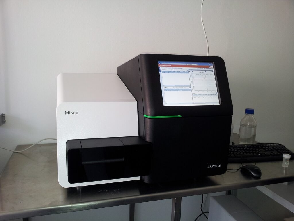
How Sequencing Data Becomes a Report
A report does not show the instrument output directly. Each region is sequenced as many short reads, which are first aligned to a reference genome, and differences between the aligned reads are then flagged as possible alterations. A large share of what is flagged at this stage is not a real change but artefact from instrument error or difficult alignment.
The raw list is therefore put through a series of filters. Regions with low read depth are set aside, variants common in the population and unrelated to disease are removed, and errors that accumulate in repetitive regions are separated out. What remains is matched to databases, named, and given a layer of clinical interpretation. The report is the list left after those filters.
Two practical points follow. The same sample run in two laboratories may not give identical reports, because panel coverage, depth thresholds and filter settings differ. And a change absent from a report has not been shown to be absent; it can only be said to fall below the detection limit of that assay. This is why an existing report is read again during a second opinion.
Panel Coverage: What a Report Cannot See
Every panel reads a defined set of genes. A fifty-gene panel and a three-hundred-gene panel do not deliver the same information. Some panels read DNA only; reliable detection of fusions requires RNA to have been analysed as well.
- How many genes the panel covers, and which.
- Whether RNA fusion analysis was performed.
- Whether copy number changes were assessed.
- Whether sequencing depth was adequate.
- Whether confirmation of germline findings was recommended.
“Nothing was found” usually means “nothing within this panel was found”. When a second opinion is sought, the coverage of the existing panel is reviewed first; a narrow panel may justify a new test. Differences in coverage are set out on the comprehensive genomic profiling page.

How an NGS Result Is Produced, Step by Step
From the journey of the sample to the laboratory to the reading of the genomic map, the video follows each step of the process.
Alterations a Report Cannot Detect
Genes outside the panel never appear, and even within covered genes certain classes of alteration cannot be detected reliably on a DNA panel. Fusions whose breakpoint falls in a non-coding part of the gene head that list; the join itself becomes visible only when it is read at RNA level.
A gene can likewise carry a normal sequence while its expression is markedly raised or reduced, which the DNA sequence does not show. Expression and fusions are pursued through RNA analysis of tumours. Reading the whole coding region instead of a panel widens coverage but lowers depth, a trade-off described under whole exome sequencing.
A sample that is inadequate for molecular work is a separate problem. Here the limit is the quality of the input rather than the breadth of the panel, and the route to take is set out under when the tissue biopsy is not enough.
How the Alterations Are Interpreted
Each line gives the gene, the type of change, its consequence at protein level and the variant allele fraction. The type tells you whether the change adds function or removes it.
- Point mutation
- A single letter changes, as in EGFR L858R.
- Insertion or deletion
- A short stretch is added or lost, as in EGFR exon 19 deletion.
- Amplification
- Copy number rises, as with HER2 amplification.
- Fusion
- Two genes join, as in ALK and ROS1 fusions.
- Deletion
- The gene is lost, common in tumour suppressors.
Each alteration carries a level of evidence. On the ESCAT scale, level I marks an alteration targeted by an approved drug in that tumour type. The level is always read with the tumour type, because the same change can sit at a different level elsewhere. The distinction is covered in driver mutation or passenger.
Send Your Reports for Assessment
Share your pathology, imaging and blood results on WhatsApp, or send them by email. Documents are reviewed before the consultation.
What TMB and MSI Mean
Tumour mutational burden counts mutations per megabase. Microsatellite instability indicates that the DNA mismatch repair system is not working. Both relate to the chance of benefit from immunotherapy.
| Finding | Typical threshold | What it means in clinic |
|---|---|---|
| High TMB | ≥10 mut/Mb, varies by panel | An immunotherapy option in some tumours |
| MSI-H / dMMR | By number of unstable markers | A strong indicator for immunotherapy |
| MSS / pMMR | Stable | This finding alone does not decide immunotherapy: no benefit is expected in colorectal cancer, but in melanoma, kidney and lung cancer immunotherapy can be standard regardless of MSS status |
| Low TMB | <10 mut/Mb | The decision rests on other findings |
Thresholds differ between panels, so TMB results from two laboratories are not directly comparable. MSI is read together with immunohistochemistry for mismatch repair proteins. The steps behind that decision are on the immunotherapy page.
Should Every Patient Have Comprehensive Profiling?
Profiling reads DNA and RNA changes, variants and copy number changes together; the video sets out when it takes priority.
Variants of Uncertain Significance
Most reports end with a long list of variants whose significance is uncertain. These lines say that current knowledge cannot tell whether the change is harmful. Treatment decisions are not built on them.
Some are reclassified over time. Laboratories update reports as evidence accumulates, so an older report can be revisited for a new question. The subject is covered in our VUS article.
The uncertainty usually comes from missing data. Calling a change harmful requires either evidence that it disrupts protein function or reports of it occurring with disease in enough people. In little-studied genes and uncommon tumours neither condition is met, so the classification stays uncertain. Uncertain does not mean harmless; it means the evidence is not yet sufficient.
Important: Whether an uncertain variant is inherited cannot be determined from a tumour panel. That question needs a separate assessment.
Common Misreadings
- Taking the number of alterations as a measure of how advanced the disease is.
- Treating a low-level finding as a reason for therapy.
- Assuming a match valid in another tumour type applies directly.
- Assuming a gene absent from the panel is intact.
- Interpreting low-fraction plasma findings without excluding clonal haematopoiesis.
- Reading the report apart from pathology and imaging.
All of these are avoided by reading the report in its clinical context. The order of assessment is described on the treatment pathway page.
Another frequent error is to apply variant explanations found online directly to one’s own report. Two different changes in the same gene can act in opposite directions; a finding that justifies treatment in one patient may mean nothing in another. Interpretation belongs with the team that knows the diagnosis and the treatment history.

What Happens After the Report Arrives
A report is not a treatment plan. It is discussed at the board; where an actionable alteration exists, approval and reimbursement are checked, and where it does not, standard treatment continues.
Recording the board decision in writing matters. If the reason a finding was set aside is not written down, the discussion starts again from nothing when the same report is reopened months later.
- The report is presented to the molecular tumour board.
- Findings are ranked by level of evidence.
- Approval, reimbursement and trial options are reviewed.
- The decision is discussed with the patient.
- At progression the report is revisited.
The limits of both markers are read on the report as well. A high mutational burden raises the chance that a tumour is visible to the immune system but does not guarantee it; where few immune cells enter the tumour, a high burden may not translate into clinical benefit. An MSI-H result shows that the repair system is not working, which can point to an inherited predisposition and needs separate assessment.
What a Genetic Panel Test Is
Two patients with the same diagnosis can have tumours that behave very differently. The video explains what the test measures.
Four questions are worth asking at the consultation: which genes did this panel cover and was RNA fusion analysis performed, does the level of evidence for the alteration apply to my tumour type, is there an approved and reimbursed drug for the finding, and does the result change my current treatment. Answered together, they make clear what the report says for this patient.
Patients should ask for and keep a copy of the report. Sharing it when a second opinion is sought usually makes repeating the same test unnecessary.
Frequently Asked Questions
How long does an NGS report take?
An NGS report takes different times depending on panel breadth and laboratory workload: a few working days for focused panels, two to three weeks for comprehensive ones. A low tumour cell fraction may force a repeat and extend the timeline.
What does it mean if no alteration is found?
A report with no alteration does not mean the tumour carries none; it means none was detectable within this panel. A narrow panel, an inadequate sample or the absence of RNA fusion analysis can all produce that result.
If TMB is high, is immunotherapy certain?
No. A high burden raises the probability of benefit but is not a decision on its own. Tumour type, PD-L1 status, MSI result and general condition are weighed together.
Does a low variant allele fraction make a finding unimportant?
Not necessarily. A low fraction may indicate a smaller subgroup of cells, but if the sample also had low tumour content the same figure reads differently. In plasma panels, low-fraction findings are additionally checked against clonal haematopoiesis.
Is the test ever repeated in the same patient?
An NGS test can be repeated. As disease progresses a tumour acquires new alterations, particularly under targeted treatment, and a fresh sample may be analysed. The earlier report is kept as well, because the comparison is useful.
Can findings in the report be passed to my family?
A tumour panel looks at changes acquired by the tumour and, as a rule, these are not inherited. Where an inherited predisposition is suspected, a separate germline test is arranged and interpreted within genetic counselling.
Should I show the report to another doctor?
Keeping a copy is advisable. Sharing it when seeking a second opinion prevents unnecessary repetition of the test and speeds up assessment.
Related Articles
Driver Mutation or Passenger: How a Genomic Report Is Read
Open the Page
What Does VUS Mean on an NGS Report?
Open the PageRelated Pages

Comprehensive Genomic Profiling
Open the PageMolecular Tumour Board
Open the PageImmunotherapy
Open the Page
Treatment Pathway
Open the PageSources
- Mosele MF, Westphalen CB, Stenzinger A et al. Recommendations for the use of next-generation sequencing (NGS) for patients with advanced cancer in 2024: ESMO Precision Medicine Working Group. Ann Oncol 2024;35(7):588-606. Link
- Mosele F, Remon J, Mateo J et al. Recommendations for the use of next-generation sequencing (NGS) for patients with metastatic cancers: ESMO Precision Medicine Working Group. Ann Oncol 2020;31(11):1491-1505. Link
- Li MM, Datto M, Duncavage EJ et al. Standards and Guidelines for the Interpretation and Reporting of Sequence Variants in Cancer (AMP/ASCO/CAP). J Mol Diagn 2017;19(1):4-23. Link
- Mateo J, Chakravarty D, Dienstmann R et al. A framework to rank genomic alterations as targets for cancer precision medicine: ESMO Scale for Clinical Actionability of molecular Targets (ESCAT). Ann Oncol 2018;29(9):1895-1902. Link
- Chakravarty D, Gao J, Phillips S et al. OncoKB: A Precision Oncology Knowledge Base. JCO Precis Oncol 2017. Link
- NCCN Clinical Practice Guidelines in Oncology, guidelines by cancer type. Link
- ESMO Clinical Practice Guidelines, guidelines by cancer type. Link