
What a Liquid Biopsy for Cancer Is
A liquid biopsy is the laboratory analysis of genetic material that cancer cells release into the bloodstream. In its most widely used form, fragments of circulating tumour DNA, known as ctDNA, are separated from a tube of blood and read with next-generation sequencing. No needle enters the tumour; the sample is collected the way any blood test is collected.
The word biopsy is misleading. The test does not provide tissue, so it cannot show how the tumour looks under a microscope, what type it is or how it is graded. What it does show is the molecular signature: which genes carry alterations, at what fraction they appear in blood, and how that picture changes over time. It therefore complements the pathology report rather than replacing it.
Three traces can be looked for in blood: cell-free ctDNA, whole circulating tumour cells and the vesicles that cells shed. In routine practice ctDNA contributes most to treatment decisions; the other two remain largely within research.
How Tumour DNA Reaches the Bloodstream
Cells inside a tumour are constantly dying. As they break apart, nuclear DNA enters the circulation in short fragments and sits in plasma alongside the cell-free DNA shed by healthy tissue. Tumour fragments are only a small share of that pool, which is where the technical difficulty lies: the signal sought resembles a far larger background. The laboratory therefore reads each region many times over, and that depth is what allows a few tumour fragments to be called reliably.
Cell-free DNA does not persist long in plasma, with two consequences in practice. The result describes the tumour at the moment of sampling, not its history, so a sample drawn weeks later can differ, and the longer the gap between sampling and the decision, the less current the report becomes. What is read from blood is also DNA, which says nothing about how actively a gene is working; expression and certain fusions are pursued through RNA analysis of tumours.
How the Test Is Done and How Long Results Take
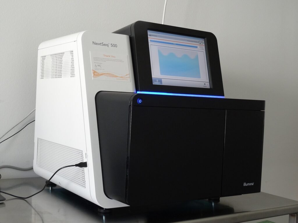
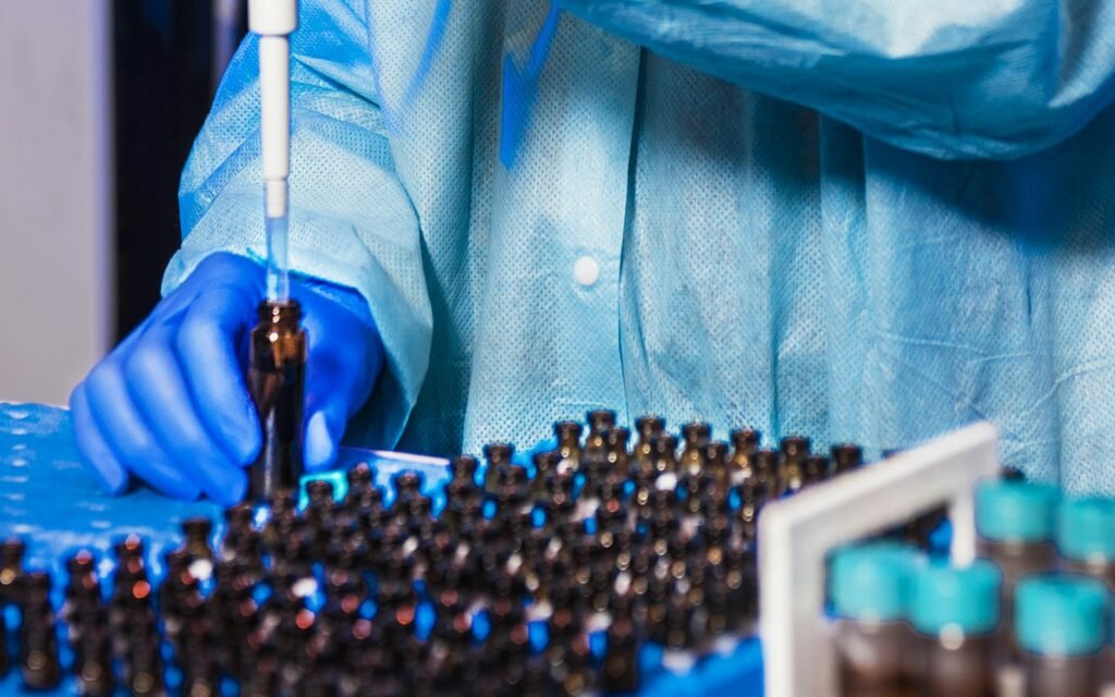
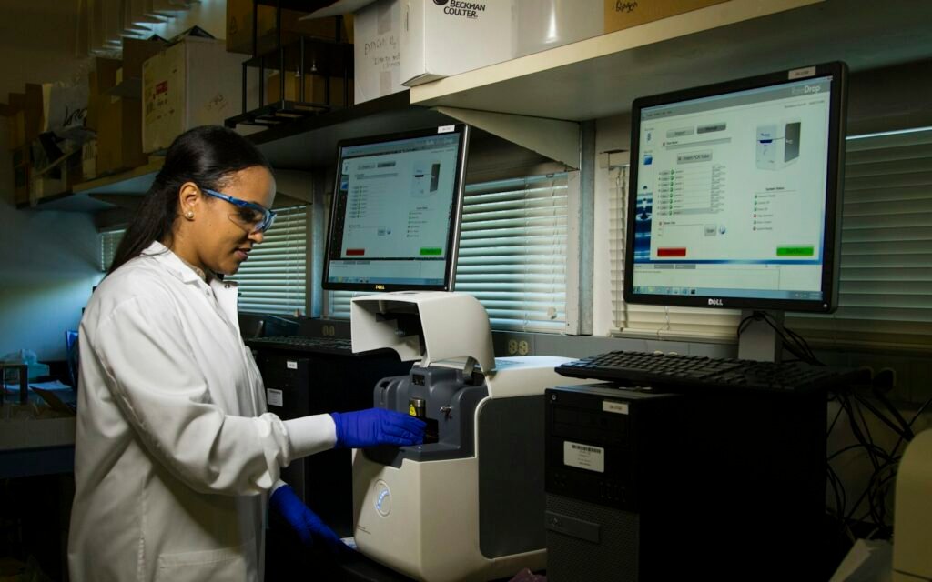
Blood is drawn into preservative tubes that slow the degradation of cell-free DNA. The volume is usually between 10 and 20 millilitres and fasting is not required. In the laboratory, plasma is separated, cell-free DNA is purified and the genes on the panel are sequenced at high depth.
- Blood is drawn into dedicated tubes and shipped to the laboratory.
- Plasma is separated and cell-free DNA is purified.
- Genes covered by the panel are sequenced at high depth.
- Detected alterations are classified by clinical significance and level of evidence.
- The report is interpreted together with the diagnosis and previous treatment.
Turnaround depends on the breadth of the panel and the workload of the laboratory: a few working days for focused panels, two to three weeks for comprehensive ones. The most common cause of delay is a low DNA yield that forces the assay to be repeated. The clinical meaning of the report is settled at the molecular tumour board.

Can a Blood Test Alone Make the Diagnosis?
Genetic tests from blood can show molecular changes in the tumour, but on their own they are not always enough to make a diagnosis. The video explains where the test fits.
When a Liquid Biopsy for Cancer Is Requested
The recommendations published by the ESMO Precision Medicine Working Group ask for plasma testing to answer a defined clinical question rather than as a routine add-on. Five situations account for most requests.
| Situation | Question asked | Effect on the decision |
|---|---|---|
| Tissue unavailable or exhausted | Is there a targetable alteration? | Targeted treatment can be started |
| A decision cannot wait | Which first-line treatment? | Treatment starts sooner |
| Progression on treatment | How did resistance arise? | The next line is chosen |
| After surgery | Is tumour DNA still detectable? | Informs the chemotherapy decision |
| Follow-up | Is the response holding? | Complements imaging |
In advanced lung cancer, when the diagnostic sample is insufficient, a plasma panel can reveal targetable alterations in EGFR, ALK, ROS1, BRAF, KRAS G12C and RET. Guidelines describe this as a plasma-first approach: if an alteration is found, treatment begins; if not, tissue is pursued again.
Why Plasma Testing Matters in Advanced Disease
Where tissue biopsy is difficult or risky, analysis from blood can guide the decision, and response and resistance can be followed the same way.
Send Your Reports for Assessment
Share your pathology, imaging and blood results on WhatsApp, or send them by email. Documents are reviewed before the consultation.
Liquid Biopsy Versus Tissue Biopsy
The two are not alternatives. Tissue establishes the diagnosis; blood describes the molecular state of the disease at that moment.
| Criterion | Tissue biopsy | Blood-based panel |
|---|---|---|
| Establishes diagnosis | Yes, the standard | No |
| Tumour type and grade | Shown | Not shown |
| Molecular alterations | Shown | Shown |
| Procedural risk | Bleeding, pneumothorax | None beyond venepuncture |
| Repeatability | Limited | Serial sampling is easy |
| Tumour heterogeneity | Reflects the sampled site | Carries traces from several sites |
| Meaning of a negative | Sample may be inadequate | Tumour may shed little DNA |
That difference matters most when a biopsy carries real risk. A deep lung lesion may bring a risk of pneumothorax; in an older patient, or one with a bleeding tendency, the procedure may be postponed. For those patients a plasma panel can supply what is needed to start treatment.
Blood or Tissue: Which Comes First?
Through the story of a 98-year-old patient with a lung mass, for whom a needle biopsy carried real risk, the video explains where each method fits.
After Surgery: Minimal Residual Disease
Once a tumour has been removed, clusters of cells too small to see may remain. This is called minimal residual disease and imaging cannot detect it. Looking for ctDNA in blood is currently the most sensitive of the methods that can show that trace.
The DYNAMIC trial in stage II colon cancer showed how far this can change practice. When adjuvant treatment was guided by the post-operative ctDNA result, chemotherapy use fell from 27.9 per cent to 15.3 per cent, and two-year recurrence-free survival was not inferior to standard management. Five-year results published later held the same shape.
Important: This does not mean that a negative result makes chemotherapy unnecessary. The decision still weighs stage, pathological risk factors, general condition and the patient’s own preference. The criteria are set out on the colorectal cancer page.
The limits of the DYNAMIC data should be stated plainly. The trial enrolled patients with stage II colon cancer, so its results cannot be carried to stage III disease, other organs or different pathological risk groups. A fall in chemotherapy use does not mean chemotherapy is unnecessary whenever no trace is detectable; what was shown is that a test-guided decision did not reduce recurrence-free survival in this group.
The same question is being studied after surgery in muscle-invasive bladder cancer, along similar lines.
The same logic is under study in bladder, breast and lung cancer. For now guidelines endorse ctDNA-guided chemotherapy decisions only in selected settings and within trials, so expectations need to be framed carefully when the test is ordered.
When Resistance Develops on Treatment
A tumour that responded well to targeted treatment can find a new escape route. The question then is why the disease is moving again, and a plasma panel can often answer it without another tissue procedure.
In lung cancer, a patient progressing on osimertinib may show an EGFR C797S alteration or MET amplification in plasma. The mechanism found directly shapes which drug comes next; the sequence is described on the targeted therapy page.
Limits and Results That Are Misread
The most misread result is a negative one. The absence of an alteration in blood does not mean the tumour lacks it; the tumour may simply not be shedding enough DNA. Shedding is low in small-volume disease, in brain-only metastases and in some tumour types.
- A negative result is confirmed on tissue whenever tissue can be obtained.
- Some alterations found in blood arise not from the tumour but from age-related clonal changes in blood cells.
- Genes outside the panel never appear in the report, so “nothing was found” also describes the panel’s limits.
- Samples taken at different times can differ; that reflects the disease, not an error in the assay.
- Inherited risk needs a separate test; a tumour panel does not answer that question.
Two misunderstandings recur. The first is that this analysis could screen healthy people; the panel described here characterises a tumour already diagnosed. The second is that every alteration has a drug behind it. For most findings there is no usable targeted agent today, which reflects the limits of the field, not a failure of the test.
Where an inherited predisposition is suspected, hereditary cancer risk assessment is planned as its own pathway. Whether an alteration seen on a tumour panel was inherited can only be settled with a confirmatory germline test.
Where the Test Sits in the Assessment Pathway
In clinic the test is not ordered on its own; it answers a question. At the first consultation the pathology report, imaging and previous treatment are reviewed, and testing is planned around whatever is missing.
When the report arrives it is read against the clinical picture. If a targetable alteration is present, approval status and reimbursement are considered; if not, standard treatment continues and tissue may be sought again. The whole sequence is set out on the treatment pathway page.
What Tissue and Blood Say Together
In cancer diagnosis, analyses from both tissue and blood are read together; a small sample reveals the genetic map and biological behaviour of the disease.
If a plasma panel has already been run elsewhere, the existing report is reviewed before a repeat is considered. Panel coverage, the date it was run and the treatment given since then determine whether testing needs to be repeated.
Frequently Asked Questions
Can a liquid biopsy for cancer make the diagnosis?
No. The test does not establish a diagnosis; it describes the molecular features of a tumour that has already been diagnosed. Diagnosis is made on pathology. Blood-based testing is used to look for targetable alterations, to identify resistance and to follow disease after treatment.
Does the procedure hurt, and is preparation needed?
Sampling is the same as any blood draw, with no additional procedure or anaesthesia, and no fasting requirement. Blood is collected into tubes that preserve cell-free DNA. Listing current medicines and previous treatment helps the report to be interpreted correctly.
What happens if the result is negative?
A negative result may mean that too little tumour DNA was circulating rather than that no alteration exists. Where tissue can be obtained, testing is repeated on tissue. Guidelines recommend returning to tissue when plasma is uninformative.
Is the test used in every cancer type?
No. It contributes most where targetable alterations are likely: lung, colorectal, breast, prostate and biliary tract cancers lead that list. Whether to request it depends on tumour type, stage and the clinical question.
Does it replace a tissue biopsy?
No. Tissue biopsy remains the standard for diagnosis and shows tumour type, grade and receptor status, none of which blood can provide. In the same patient the two answer different questions and are often used together.
How long do results take?
Focused panels report within a few working days; comprehensive panels take two to three weeks. Timing depends on laboratory workload, panel breadth and the amount of DNA recovered. A repeat assay after a low yield extends it.
Does it show inherited cancer risk?
A blood or tumour panel does not show inherited risk. A tumour panel looks for alterations acquired by the tumour. Inherited predisposition requires a separate germline test, interpreted within genetic counselling.
Related Articles
What Does VUS Mean on an NGS Report?
Open the Page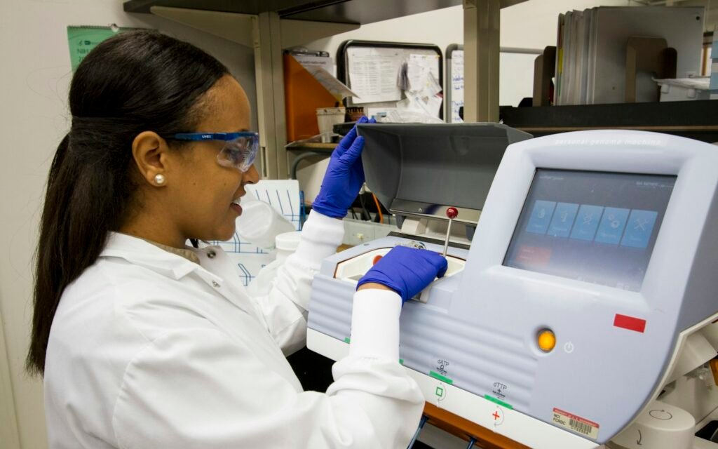
Why Treatment Sequencing in Metastatic Colorectal Cancer Starts With Biomarkers
Open the PageRelated Pages
Precision Oncology
Open the PageComprehensive Genomic Profiling
Open the Page
Molecular Tumour Board
Open the Page
Treatment Pathway
Open the PageSources
- Pascual J, Attard G, Bidard F-C et al. ESMO recommendations on the use of circulating tumour DNA assays for patients with cancer. Ann Oncol 2022;33(8):750-768. Link
- Tie J, Cohen JD, Lahouel K et al. Circulating tumor DNA analysis guiding adjuvant therapy in stage II colon cancer (DYNAMIC). N Engl J Med 2022;386:2261-2272. Link
- Tie J et al. Circulating tumor DNA analysis guiding adjuvant therapy in stage II colon cancer: 5-year outcomes of the randomized DYNAMIC trial. Nat Med 2025. Link
- NCCN Clinical Practice Guidelines in Oncology: Non-Small Cell Lung Cancer, biomarker testing section. Link
- U.S. Food and Drug Administration. List of cleared or approved companion diagnostic devices (in vitro and imaging tools). Link
- ESMO Precision Medicine Working Group. The use of circulating tumour DNA assays for patients with cancer (OncologyPRO summary). Link