
What BCC Is and Who Is Affected
Basal cell carcinoma (BCC) is a cancer that arises from the basal cells at the bottom of the epidermis, the outer layer of the skin, and it is the most common cancer in humans. It makes up about 75 per cent of all skin cancers; IARC records more than a million non-melanoma skin cancers a year worldwide, and most of them are BCC. The tumour grows slowly over years and invades the surrounding tissue, but spread to the lymph nodes or distant organs occurs in fewer than 0.1 per cent of cases.
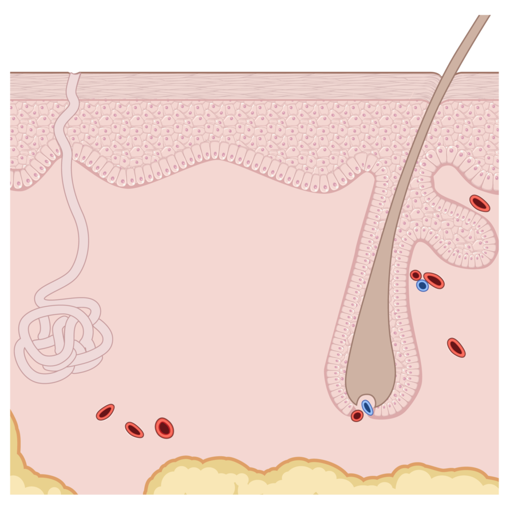
BCC is most common over the age of 60 in fair-skinned people with a lifetime of sun exposure; about 80 per cent of tumours are on the head and neck, particularly the nose, cheeks, forehead and ears. Smoking, previously irradiated skin, arsenic exposure and immunosuppressive drugs after organ transplantation raise the risk. The other types of skin cancer are compared on the skin cancers page.
Gorlin syndrome (basal cell naevus syndrome) is considered in people who develop many BCCs at a young age; it is caused by an inherited (germline) mutation in the PTCH1 gene and is accompanied by jaw cysts and small pits on the palms. People with Gorlin syndrome need lifelong dermatology follow-up. There is no screening programme for BCC in the general population; anyone with a non-healing sore or a new shiny bump should see a dermatologist.
Appearance and Subtypes
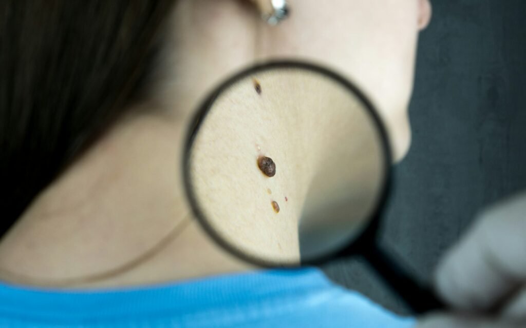
The typical BCC is a shiny or pearly bump with fine visible blood vessels that grows slowly over months or years on sun-exposed skin. The centre may sink and ulcerate; it crusts, bleeds, seems to heal and then opens again, and it is usually painless. The appearance depends on the subtype, and the subtype influences the choice of treatment; the pathologist makes this distinction on the biopsy.
- Nodular BCC
- The most common subtype (about 60 per cent): a dome-shaped, shiny, pink or skin-coloured bump on the face with fine red vessels on its surface. As it grows the centre sinks and ulcerates. It is treated by surgical excision.
- Superficial BCC
- A pink, slightly scaly, flat, well-defined patch on the trunk, arms or legs that can be mistaken for eczema or a fungal infection. Because it does not grow deep into the skin, non-surgical options such as creams, cryotherapy and photodynamic therapy are suitable for this subtype.
- Morphoeic (sclerosing) BCC
- A firm, whitish or yellowish, scar-like area with indistinct edges that extends beyond what is visible, so diagnosis and complete removal are difficult. It is a high-risk subtype that needs Mohs surgery or wide excision, and its recurrence rate is higher than that of the other subtypes.
- Pigmented BCC
- A nodular or superficial BCC containing brown or black pigment. It can be confused with melanoma and is distinguished by dermoscopy; if melanoma is suspected, the criteria on the melanoma page apply.
Diagnosis and Staging
BCC is diagnosed by dermoscopy (examination with a magnifying, illuminated instrument) followed by pathological examination of a punch, shave or excisional biopsy; the report states the subtype, depth and whether the tumour has grown around nerves (perineural invasion). No blood tests or imaging are needed for diagnosis; CT or MRI is requested only for large, deep tumours where bone, orbit or nerve involvement is suspected.
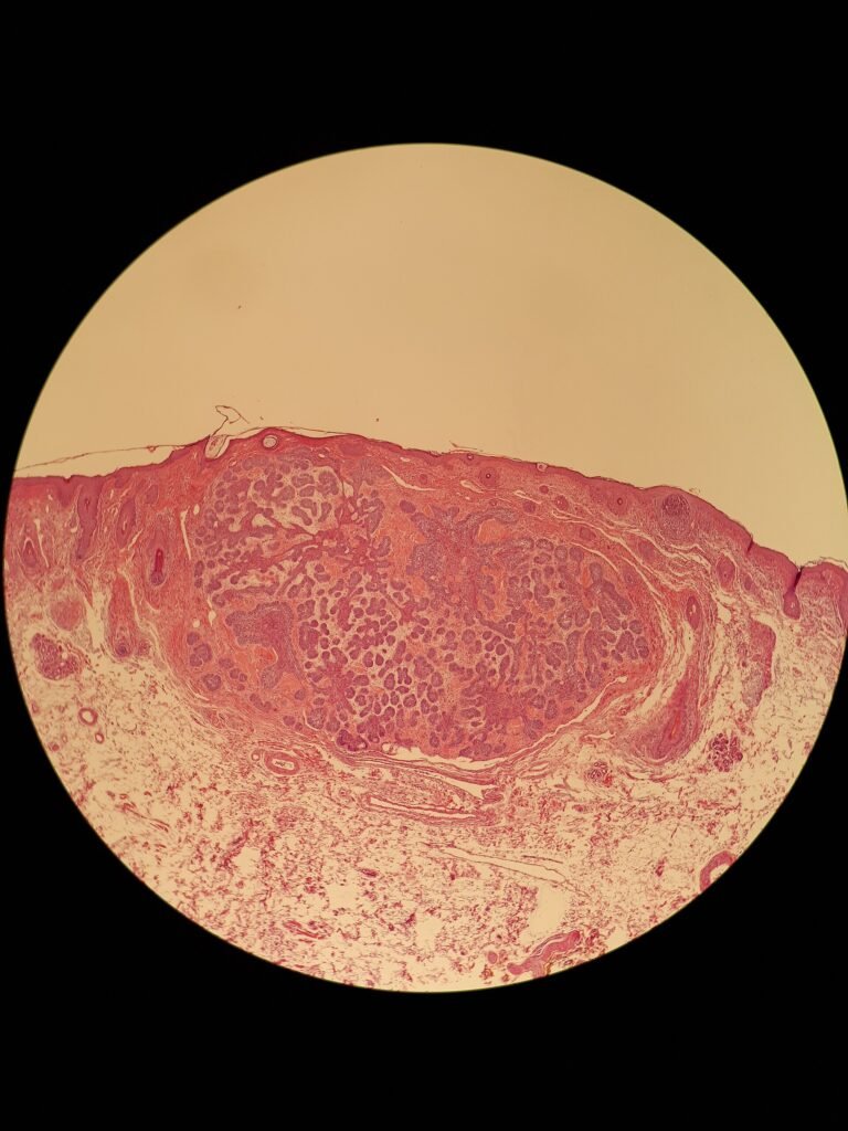
Rather than the classic TNM stage, BCC is classified by the risk categories in the NCCN and European (EADO/EDF/EORTC 2023) guidelines, because almost all tumours present without lymph node or distant spread. Low risk: a first tumour under 2 cm on the trunk or limbs, nodular or superficial, with well-defined edges. High risk: a site on the face, ears, hands, feet or genital area; indistinct edges; morphoeic or infiltrative subtype; perineural invasion; recurrent tumour; previously irradiated skin; and immunosuppression. Treatment is selected by the treating clinician according to this risk category and the site of the tumour.
Locally advanced BCC describes a large, deep, recurrent tumour, or one that has grown into bone or the orbit, for which surgery or radiotherapy is not possible or would cause unacceptable loss of function or appearance; this judgement is made at a meeting of the surgeon, radiation oncologist and medical oncologist. Metastatic BCC covers the very rare cases that have spread to lymph nodes, lungs or bone. These two groups are the subject of medical oncology treatment. Whether a tumour counts as locally advanced can usually be judged remotely from the pathology report, clinical photographs and any CT or MRI, and the steps for sending them are described on the treatment pathway page.
Surgery and Local Treatments
The large majority of BCCs are removed under local anaesthetic by dermatology, plastic surgery or dermatological surgery and do not need a medical oncology opinion. In standard excision the tumour is removed with a 4 mm margin of healthy skin and the pathologist confirms that the margin is clear; the five-year recurrence rate for low-risk tumours is under 5 per cent. Mohs micrographic surgery examines the margins of the removed tissue layer by layer under the microscope during the operation; it gives the lowest recurrence rate for high-risk tumours and for sites such as the face where preserving tissue matters. Reconstruction is planned by plastic surgery.
| Treatment | Where it is used | Specialty |
|---|---|---|
| Standard surgical excision | The large majority of low- and intermediate-risk tumours; 4 mm margin | Dermatology, plastic surgery |
| Mohs micrographic surgery | Face, periocular area, nose and ears; morphoeic and recurrent tumours | Dermatological surgery |
| Radiotherapy | Older patients who decline or are unfit for surgery; after surgery when margins are positive or perineural invasion is present | Radiation oncology |
| Curettage and electrodesiccation, cryotherapy | Small, low-risk nodular or superficial tumours on the trunk and limbs | Dermatology |
| Topical imiquimod or 5-fluorouracil, photodynamic therapy | Superficial subtype only, on low-risk sites, when a higher recurrence rate than surgery is accepted | Dermatology |
| Hedgehog pathway inhibitor, immunotherapy | Locally advanced or metastatic tumour when surgery and radiotherapy are unsuitable | Medical oncology |
Radiotherapy gives control close to that of surgery in older patients who cannot have or do not want an operation, but it is not the first choice in younger patients because of the long-term risk of new skin cancers and tissue changes in the treated area, and it is not used in Gorlin syndrome. Treatment is selected by the treating clinician according to risk category, pathology and site, and the patient's expectations regarding appearance and function are part of the decision.
Important: The information on this page describes general principles. It does not replace a consultation or an individual treatment plan. For the large majority of basal cell carcinomas the first point of contact is a dermatologist; a medical oncology opinion is needed for locally advanced and metastatic disease. Licensing and access to specific drugs are discussed at the consultation as they stand on 21 September 2026.
Systemic Therapy for Locally Advanced and Metastatic BCC
Systemic therapy (drug treatment that acts throughout the body) is needed in BCC only for locally advanced tumours that cannot be controlled by surgery and radiotherapy and for metastatic disease; together these make up less than 1 per cent of all BCCs. First-line treatment is a hedgehog pathway inhibitor: about 90 per cent of BCCs carry a mutation in PTCH1 or SMO that keeps the hedgehog signalling pathway permanently switched on, and the tumour depends on this pathway to grow. Vismodegib (ERIVANCE trial) and sonidegib (BOLT trial) are oral targeted drugs taken once daily that block the pathway at the SMO protein; in locally advanced disease they shrink the tumour in about 45 to 60 per cent of patients, and the response is usually seen within two to three months.
Side effects of hedgehog inhibitors occur in most patients and determine how well treatment is tolerated: muscle cramps (about two thirds of patients), loss or distortion of taste, hair loss, weight loss, tiredness and nausea. These effects reverse when the drug is stopped, and they are managed by intermittent dosing or planned treatment breaks. Because the drug causes severe developmental defects in an unborn child, effective contraception is mandatory during treatment and for 24 months after vismodegib or 20 months after sonidegib, and blood donation is not allowed. The principles of this drug class are explained on the targeted therapy page.
For patients whose disease progresses on a hedgehog inhibitor, or who cannot tolerate it, the second-line treatment is the PD-1 inhibitor cemiplimab; in its phase 2 trial about 30 per cent of patients with locally advanced disease had tumour shrinkage, and responses were durable. It is given intravenously every three weeks, and immune-related side effects (thyroid dysfunction, colitis, rash, pneumonitis) are monitored; the general principles are described on the immunotherapy page. Using a hedgehog inhibitor before surgery to shrink a tumour, or to prevent new tumours in Gorlin syndrome, is a matter for assessment in selected patients; platinum chemotherapy is used only rarely when other options are exhausted.
Oral Therapy for a Patient from Abroad
Because hedgehog inhibitors are taken by mouth, a patient from abroad does not need to stay in Izmir for the whole course. After the first consultation, the start of treatment and the first response check at two to three months, follow-up can continue with the home physician using a written plan that sets out the dose, the side effects to watch for and the timing of clinical photographs or imaging. Vismodegib, sonidegib and cemiplimab are licensed in Türkiye; access arrangements for a visiting patient are confirmed before treatment starts.
Recurrence and Follow-Up
Follow-up after BCC treatment has two aims: to catch recurrence at the same site early and to find new tumours. The chance of a treated BCC recurring at the same site is under 5 per cent for a low-risk tumour and higher for high-risk tumours and positive margins; most recurrences appear within the first three years. Because about 40 per cent of people who have had one BCC develop another within five years, the emphasis of follow-up shifts to looking for new lesions; these people also have a higher risk of squamous cell carcinoma and melanoma.
NCCN and European guidelines recommend a full skin examination every six months in the first year, then yearly for at least five years; with multiple or high-risk tumours, immunosuppression or Gorlin syndrome, checks are every three to six months for life. Patients on a hedgehog inhibitor or immunotherapy are reviewed by medical oncology for side effects at every cycle and for response every two to three months by imaging or measured clinical photographs. Patients check their own skin monthly with a mirror and report a new non-healing sore, a lump in the scar or bleeding without waiting for the next appointment.
Sun protection is part of follow-up: a broad-spectrum SPF 30 or higher sunscreen, a hat and clothing, shade between 11:00 and 16:00 and avoiding sunbeds reduce the risk of new tumours. In the ONTRAC trial, nicotinamide (vitamin B3) reduced the number of new skin cancers in high-risk people; its use is discussed with the treating physician. To request a medical oncology assessment for locally advanced or metastatic BCC, the routes on the contact page can be used; the pathology report, clinical photographs and any imaging are reviewed before the consultation, and correspondence is in English.
Frequently Asked Questions
Is basal cell carcinoma dangerous?
Basal cell carcinoma very rarely threatens life. Because it does not spread to distant organs, the large majority of patients are treated permanently with surgery or local methods. The risk arises in tumours left untreated for years that have grown into the eye, nose, bone or skull, and in the metastatic disease seen in fewer than 0.1 per cent of cases. Even then, hedgehog inhibitors and immunotherapy can bring the disease under control. The outcome depends on the size and site of the tumour and varies from person to person.
Does BCC spread?
Basal cell carcinoma spreads to the lymph nodes or distant organs only very rarely; European guidelines put the metastasis rate below 0.1 per cent. Its real characteristic is local growth that destroys skin, cartilage and bone, which is why tumours left untreated for a long time on the face, around the eye or on the nose cause problems. The rare metastatic cases are usually very large, deep, recurrent or long-neglected tumours. In these cases a medical oncologist plans systemic treatment.
What happens if it is not treated?
An untreated basal cell carcinoma does not go away on its own. Over years it enlarges, ulcerates and bleeds, and it destroys neighbouring skin, muscle, cartilage and bone. On the eyelid, nose and ear this progression can cause loss of sight, permanent disfigurement and the need for major reconstructive surgery. Invasion of the skull and nerves is rare but serious. Because most tumours treated early are removed with a small operation and little scarring, a suspicious lesion should be shown to a dermatologist promptly.
Can it be treated with cream?
Yes, but only for superficial basal cell carcinomas that are small and on low-risk sites such as the trunk, arms and legs. Imiquimod cream is applied five days a week for six weeks and 5-fluorouracil cream twice daily for four weeks; the treated area becomes red, crusted and sore. The recurrence rate after cream is higher than after surgery, so cream is not used for nodular, morphoeic, facial or recurrent tumours. The dermatologist decides on the basis of the biopsy result.
What is a hedgehog inhibitor?
A hedgehog inhibitor is an oral targeted drug that blocks the hedgehog signalling pathway on which basal cell carcinoma depends for growth; vismodegib and sonidegib belong to this class. A medical oncologist prescribes them for locally advanced or metastatic tumours that are unsuitable for surgery and radiotherapy, and they shrink the tumour in about 45 to 60 per cent of patients. The most common side effects are muscle cramps, loss of taste, hair loss, weight loss and tiredness. Contraception is mandatory during and after treatment.
How often are check-ups needed?
After treatment of a basal cell carcinoma, a dermatology check is done every six months in the first year and then yearly for at least five years. People who have had several tumours or a high-risk tumour, and people with Gorlin syndrome, are checked every three to six months for life. About 40 per cent of people who have had one basal cell carcinoma develop another within five years, so follow-up is aimed more at finding new lesions than at recurrence. Patients also check their own skin monthly.
Related Pages
Skin Cancers
Open the Page
Melanoma
Open the Page
Targeted Therapy
Open the Page
Immunotherapy
Open the Page
Treatment Pathway
Open the Page
Contact
Open the Page
International Patients
Open the PageSources and Guidelines
- Peris K et al. European consensus-based interdisciplinary guideline for diagnosis and treatment of basal cell carcinoma, update 2023 (EADO, EDF, EORTC). Eur J Cancer 2023;192:113254. Link
- National Comprehensive Cancer Network. NCCN Clinical Practice Guidelines in Oncology: Basal Cell Skin Cancer. Link
- Sekulic A et al. Efficacy and safety of vismodegib in advanced basal-cell carcinoma (ERIVANCE BCC). N Engl J Med 2012;366:2171-2179. Link
- Migden MR et al. Treatment with two different doses of sonidegib in patients with locally advanced or metastatic basal cell carcinoma (BOLT). Lancet Oncol 2015;16(6):716-728. Link
- Stratigos AJ et al. Cemiplimab in locally advanced basal cell carcinoma after hedgehog inhibitor therapy: an open-label, multi-centre, single-arm, phase 2 trial. Lancet Oncol 2021;22(6):848-857. Link
- Chen AC et al. A phase 3 randomized trial of nicotinamide for skin-cancer chemoprevention (ONTRAC). N Engl J Med 2015;373:1618-1626. Link
- National Cancer Institute. Skin Cancer Treatment (PDQ), patient version. Link
- Cancer Research UK. Basal cell skin cancer. Link
- International Agency for Research on Cancer (WHO). GLOBOCAN 2022, Global Cancer Observatory. Link